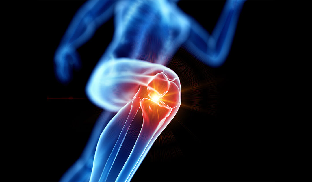
Even Light Exercise Can Help Arthritic Knees

Even Light Exercise Can Help Arthritic Knees

Even short bouts of light exercise can help the millions of people with knee osteoarthritis reduce pain and improve their range of motion.

Knee osteoarthritis, the wear-and-tear form of the disease, occurs when the cartilage between your bones breaks down, causing pain, stiffness and swelling.

Researchers in a new study compared high-dose and low-dose exercise in 189 people with knee osteoarthritis. Everyone exercised three times a week for 12 weeks. Exercises included indoor cycling and various lower body exercises such as de-loaded squats and knee extensions. De-loaded exercises use bands or pulleys to take weight off the knee joint and minimize pain.

Folks in the high-dose group performed 11 exercises in 60- to 90-minute sessions. Folks in the low-dose exercise group performed five exercises during 20- to 30-minute sessions.

The result? Everyone showed improvements on a standard scale measuring knee osteoarthrosis pain and function at three, six and 12 months. People in the high-dose group did show greater improvements in knee function during sports and recreation at six months, suggesting that a high-dose program may be better for athletes and weekend warriors.

The study, led by Wilhelmus Johannes Andreas Grooten, a physiotherapist at the Karolinska Institute in Stockholm, was published Jan. 24 online in the Annals of Internal Medicine.

Outside experts are quick to point out that the best exercise plan for someone with knee osteoarthritis should fit in with their lifestyle, goals and capabilities.

Jack Fitzgerald is a physical therapist at the Hospital for Special Surgery in New York City. He routinely designs and implements exercise plans for people with knee osteoarthritis.

Calling the new study results “music to any physical therapist’s ears,” Fitzgerald pointed out that participants in the study showed meaningful improvements in knee symptoms and that adherence to their program was excellent across both groups.

This is at least partially due to the fact that exercise was prescribed on a minimal/no pain basis, Fitzgerald said.

“Regardless of the exercise dosage, when we are capable of prescribing exercise within pain-free limits, there is more hope that patients will respond well to the treatment,” he said.

“If you are prescribing exercises for these patients with their best interest in mind, the program should be specific to the likes and goals of the patient keeping in mind loading principles to manage pain,” Fitzgerald added.

Exercise is an essential part of knee osteoarthritis treatment, said Dr. Sonali Khandelwal, an associate professor of internal medicine at Rush Medical College in Chicago.

“A component of knee osteoarthritis is weakening of the muscles surrounding the knee, and exercise strengthens these supporting muscles,” she said.

Many people worry that exercise will make knee OA worse, but this isn’t true, Khandelwal said.

“It’s important to assess a person’s risk of falls and history of exercise when prescribing a plan,” she said. “For people who have never done any exercise, just moving and walking tends to go a long way.”

Conservative measures such as physical therapy and exercise are first-line treatments for knee osteoarthritis pain, Khandelwal said. Next up are steroid or hyaluronic gel injections and pain-relieving medication like non-steroidal anti-inflammatory drugs or acetaminophen.

“When conservative measures don’t work, pain is a 10 out of 10, and you can’t perform activities of daily living, surgery can be considered,” she said.

“It’s great to see studies like this as surgery results don’t last forever,” Khandelwal said.

Dr. David Pisetsky, a professor of medicine at Duke University School of Medicine in Durham, N.C., agreed.

“For people with knee osteoarthritis, activity is important to maintain strength and range of motion,” he said. “Exercise should be easy to perform … walking is a good way to go.”

Other options include swimming, riding a stationary bicycle, and exercising with light weights.

More information

The Arthritis Foundation provides more on knee osteoarthritis, including its causes and treatments.

SOURCES: Jack Fitzgerald, DPT, physical therapist, Hospital for Special Surgery, New York City; Sonali Khandelwal, MD, associate professor, internal medicine, Rush Medical College, Chicago; David Pisetsky, MD, PhD, professor, medicine, Duke University School of Medicine, Durham, N.C; Annals of Internal Medicine, Jan. 24, 2023

Jog on: Exercise Won’t Raise Your Odds for Arthritic Knees

Jog on: Exercise Won’t Raise Your Odds for Arthritic Knees

Dr. Kim Huffman, an avid runner, gets a fair amount of guff from friends about the impact that her favorite exercise has on her body.

“People all the time tell me, ‘Oh, you wait until you’re 60. Your knees are going to hate you for it’,” Huffman said. “And I’m like, ‘That’s ridiculous’.”

Next time the topic comes up, Huffman is well-armed: An extensive British analysis of prior study data has found no link between a person’s amount of exercise and their risk for knee arthritis.

The research team combined the results of six clinical trials conducted at different places around the globe, creating a pool of more than 5,000 people who were followed for 5 to 12 years for signs of knee arthritis.

In each clinical trial, researchers tracked participants’ daily activities and estimated the amount of energy they expended in physical exertion.

Neither the amount of energy burned during exercise nor the amount of time spent in physical activity had anything to do with knee pain or arthritis symptoms, the researchers concluded.

“This helps dispel a myth that I’ve been trying to dispel for quite a while,” said Huffman, an associate professor at the Duke University Medical Center’s division of rheumatology.

“If you add up the amounts of activity that people do and also the duration of activity, neither of those is associated with knee arthritis,” added Huffman, who wasn’t involved in the analysis.

Dr. Bert Mandelbaum is chief medical officer of the Los Angeles Galaxy soccer club and team physician for the U.S. Soccer Men’s National Team.

He agreed the study “further corroborates the fact that levels of exercise in one’s personal life do not increase the risk, the onset or progression of osteoarthritis.”

So where did this misconception come from?

Huffman thinks it’s because people mistake exercise-related injuries for the effect that exercise itself has on your joints.

“Right now, the clear risks for knee arthritis are genetics, injuries and female sex,” Huffman said. “People who exercise more may be more likely to injure their knee. That’s where I think the myth comes from.”

In fact, exercise can help ward off knee arthritis in several ways, Huffman said:

Flexing and extending the knee during exercise promotes the diffusion of fluid into the joint, promoting better nutrition.

An elevated metabolism created by exercise helps control inflammation in the knee joint.

Weight loss reduces the amount of load placed on the knee.

Exercise strengthens the muscles surrounding the knee, stabilizing it and reducing the risk of injury.

“I don’t think we’re finding that simple overuse or using your joint is a problem. It’s more an association with injuries and perhaps in the setting of obesity or high genetic risk,” Huffman said.

Your best bet is to choose an exercise that poses the least risk of a knee injury, Huffman said.

“If you want to go snow skiing, I don’t think that’s a huge problem but you’re probably going to be more likely to injure yourself downhill skiing than, say, walking in your neighborhood or training for a marathon,” Huffman said. “It’s not soccer or football or skiing itself. It’s just the risk for injury during those activities.”

On the other hand, exercise provides benefits that go far beyond healthy joints, said Mandelbaum, co-chair of medical affairs at Cedars-Sinai Kerlan-Jobe Institute at Santa Monica, Calif. He played no role in the research review.

“Physical activity is essential to optimize both physical and mental health and plays a central role in facilitating life’s quality and quantity,” Mandelbaum said. “The list of benefits includes decreased anxiety, better mood, decreased levels of coronary disease, hypertension, diabetes and obesity, and therefore a longer life.”

The analysis was published recently in the journal Arthritis and Rheumatology.

More information

The Arthritis Foundation has more about knee osteoarthritis.


SOURCES: Kim Huffman, MD, PhD, associate professor, Duke University Medical Center, division of rheumatology; Bert Mandelbaum, MD, co-chair, medical affairs, Cedars-Sinai Kerlan-Jobe Institute, Santa Monica, Calif.; Arthritis and Rheumatology, Nov. 3, 2021

What to Do If You Wake Up in Pain

What to Do If You Wake Up in Pain

Feeling achy and stiff in the morning? Try these seven techniques to ease into the day.

Nothing is more restorative than a good night’s sleep. You wake up refreshed and ready to take on a new day. Yet, for some people, the early morning hours bring unwelcome neck and back pain.

Movement during the day promotes fluid secretion from your tissues, which in turn keeps your joints mobile. When you’re asleep, however, you’re not moving for an extended period of time, which can cause your joints and muscles to stiffen up.

For some people, morning pain and stiffness go away as the day goes on. For others, the pain lasts all day. If you find yourself waking up constantly in pain, try these seven tips for a pain-free morning:

1. Stretch

When you wake up, do some simple stretches to unlock your joints and muscles. Here are two to try:

  • Knee to Chest Stretch: Remain on your back in your bed, and pull both knees toward you until your feet are flat on the bed. Grab your right knee with both hands and pull toward your chest. Do the same with the left knee. Hold for about 30 seconds. Repeat two to three times.
  • Shoulder/Upper Back Stretch: Stand up and hold your right elbow with your left hand. Stretch your right arm across the front of your body, keeping your arm straight. Do the same with your opposite arm. Hold for 30 seconds, and do two to three repetitions.

2. Change Your Mattress and Pillow

Your morning pain and stiffness could be caused by your bed or pillow. Be sure to choose a pillow that aligns your neck parallel to the mattress so it isn’t bent up or down. As for your mattress, it should be firm, but not too firm.

3. Change Your Sleep Position

Sleeping on your stomach may be the root of your pain. If so, switch to your back and prop up your knees with a pillow to keep your spine in a neutral position. Side-sleepers can try inserting a pillow between their knees.

4. Exercise Regularly

An intense workout just before you slip under the covers is not advisable. However, a brisk walk or relaxing yoga during the day can loosen up your muscles and joints, setting you up for a pain-free morning. Typically, thirty minutes of exercise a day will tire you out so you can fall asleep faster.

5. Stay Hydrated

During the night, you lose water when you sweat, which can lead to dehydration. Dehydration stiffens your muscles and joints as your tissues are deprived of fluids. So instead of going for that cup of morning coffee, drink two glasses of water first to rehydrate your body.

6. Take Vitamin D

Most people don’t get enough vitamin D in their diet. But the vitamin is essential for bone and muscle health. Have your doctor test your vitamin D levels, and if you are deficient, take a vitamin D supplement regularly.

7. Visit a Physical Therapist

Lingering morning pain should be checked out by a physical therapist. Keep a log of where the pain is strongest and which actions seem to aggravate it. Your physical therapist can show you how to adjust your sleeping position to prevent pain and improve your posture. He or she can also recommend the right pillow and mattress for a restful night’s slumber.

Don’t Live With Pain

If you’re experiencing pain and stiffness in the morning, the orthopedic specialists at Comprehensive Orthopaedics can review your symptoms to determine the source of your discomfort.

Tennis Elbow (Lateral Epicondylitis)

Tennis Elbow (Lateral Epicondylitis)

Tennis elbow, or lateral epicondylitis, is a painful condition of the elbow caused by overuse. Not surprisingly, playing tennis or other racquet sports can cause this condition. However, several other sports and activities besides sports can also put you at risk.  Tennis elbow is inflammation or, in some cases, microtearing of the tendons that join the forearm muscles on the outside of the elbow. The forearm muscles and tendons become damaged from overuse — repeating the same motions again and again. This leads to pain and tenderness on the outside of the elbow.There are many treatment options for tennis elbow. In most cases, treatment involves a team approach. Primary doctors, physical therapists and, in some cases, surgeons work together to provide the most effective care.

Anatomy

Your elbow joint is a joint made up of three bones: your upper arm bone (humerus) and the two bones in your forearm (radius and ulna). There are bony bumps at the bottom of the humerus called epicondyles, where several muscles of the forearm begin their course. The bony bump on the outside (lateral side) of the elbow is called the lateral epicondyle.

ECRB muscle and tendon

The ECRB muscle and tendon is usually involved in tennis elbow.
Reproduced and modified from The Body Almanac. © American Academy of Orthopaedic Surgeons, 2003.

Muscles, ligaments, and tendons hold the elbow joint together.

Lateral epicondylitis, or tennis elbow, involves the muscles and tendons of your forearm that are responsible for the extension of your wrist and fingers. Your forearm muscles extend your wrist and fingers. Your forearm tendons — often called extensors — attach the muscles to bone.  The tendon usually involved in tennis elbow is called the Extensor Carpi Radialis Brevis (ECRB).

Cause

Overuse

Recent studies show that tennis elbow is often due to damage to a specific forearm muscle. The extensor carpi radialis brevis (ECRB) muscle helps stabilize the wrist when the elbow is straight. This occurs during a tennis groundstroke, for example. When the ECRB is weakened from overuse, microscopic tears form in the tendon where it attaches to the lateral epicondyle. This leads to inflammation and pain.

The ECRB may also be at increased risk for damage because of its position. As the elbow bends and straightens, the muscle rubs against bony bumps. This can cause gradual wear and tear of the muscle over time.

Activities

Athletes are not the only people who get tennis elbow. Many people with tennis elbow participate in work or recreational activities that require repetitive and vigorous use of the forearm muscle or repetitive extension of the wrist and hand.

Painters, plumbers, and carpenters are particularly prone to developing tennis elbow. Studies have shown that auto workers, cooks, and even butchers get tennis elbow more often than the rest of the population. It is thought that the repetition and weight lifting required in these occupations leads to injury.

Tennis player

Playing tennis is a possible cause of tennis elbow, but other activities can also put you at risk.

Thinkstock © 2015.

Age

Most people who get tennis elbow are between the ages of 30 and 50, although anyone can get tennis elbow if they have the risk factors. In racquet sports like tennis, improper stroke technique and improper equipment may be risk factors.

Unknown

Lateral epicondylitis can occur without any recognized repetitive injury. This occurrence is called “idiopathic” or of an unknown cause.

Symptoms

The symptoms of tennis elbow develop gradually. In most cases, the pain begins as mild and slowly worsens over weeks and months. There is usually no specific injury associated with the start of symptoms.

Common signs and symptoms of tennis elbow include:

  • Pain or burning on the outer part of your elbow
  • Weak grip strength
  • Sometimes, pain at night

The symptoms are often worsened with forearm activity, such as holding a racquet, turning a wrench, or shaking hands. Your dominant arm is most often affected; however, both arms can be affected.

Location of pain in lateral epicondylitis.

Location of pain in lateral epicondylitis.

Doctor Examination

Your doctor will consider many factors in making a diagnosis. These include how your symptoms developed, any occupational risk factors, and recreational sports participation.

Your doctor will talk to you about what activities cause symptoms and where on your arm the symptoms occur. Be sure to tell your doctor if you have ever injured your elbow. If you have a history of rheumatoid arthritis or nerve disease, tell your doctor.

During the examination, your doctor will use a variety of tests to pinpoint the diagnosis. For example, your doctor may ask you to try to straighten your wrist and fingers against resistance with your arm fully straight to see if this causes pain. If the tests are positive, it tells your doctor that those muscles may not be healthy.

doctor pressing lateral epicondyle

During the exam, your doctor will apply gentle pressure to the lateral epicondyle, checking for pain and tenderness.

Tests

Your doctor may recommend additional tests to rule out other causes of your problem.

  • X-rays. These tests provide clear images of dense structures, such as bone. They may be taken to rule out arthritis of the elbow.
  • Magnetic resonance imaging (MRI) scan.  MRI provides images of the body’s soft tissues, including muscles and tendons. An MRI scan may be ordered to determine the extent of damage in the tendon or to rule out other injuries. If your doctor thinks your symptoms might be related to a neck problem, he or she may order an MRI scan of the neck to see if you have a herniated disk or arthritic changes in your neck. Both of these conditions can produce arm pain.
  • Electromyography (EMG). Your doctor may order an EMG to rule out nerve compression. Many nerves travel around the elbow, and the symptoms of nerve compression are similar to those of tennis elbow.

Treatment

Nonsurgical Treatment

Approximately 80% to 95% of patients have success with nonsurgical treatment.

Rest. The first step toward recovery is to give your arm proper rest. This means that you will have to stop or decrease participation in sports, heavy work activities, and other activities that cause painful symptoms for several weeks.

Medications. Acetaminophen or anti-inflammatory medications (such as ibuprofen) may be taken to help reduce pain and swelling

Physical therapy. Specific exercises are helpful for strengthening the muscles of the forearm. Your therapist may also perform ultrasound, ice massage, or muscle-stimulation techniques to improve muscle healing.

Wrist stretching exercise

Wrist stretching exercise with elbow extended.

Brace. Using a brace centered over the back of your forearm may also help relieve symptoms of tennis elbow. This can reduce symptoms by resting the muscles and tendons.

Counterforce brace

Counterforce brace.

Steroid injections. Steroids, such as cortisone, are very effective anti-inflammatory medicines. Your doctor may decide to inject the painful area around your lateral epicondyle with a steroid to relieve your symptoms.

Platelet-rich plasma. Platelet-rich plasma (PRP) is a biological treatment designed to improve the biologic environment of the tissue. This involves obtaining a small sample of blood from the arm and centrifuging it (spinning it) to obtain platelets from the solution.  Platelets are known for their high concentration of growth factors, which can be injected into the affected area. While some studies about the effectiveness of PRP have been inconclusive, others have shown promising results.

PRP injection

An injection of PRP is used to treat tennis elbow.
Courtesy of Allan K. Mishra, MD, Menlo Park, CA.

Extracorporeal shock wave therapy. Shock wave therapy sends sound waves to the elbow. These sound waves create “microtrauma” that promotes the body’s natural healing processes. Shock wave therapy is considered experimental by many doctors, but some sources show it can be effective.

Equipment check. If you participate in a racquet sport, your doctor may encourage you to have your equipment checked for proper fit. Stiffer racquets and looser-strung racquets often can reduce the stress on the forearm, which means that the forearm muscles do not have to work as hard. If you use an oversized racquet, changing to a smaller head may help prevent symptoms from recurring.

Surgical Treatment

If your symptoms do not respond after 6 to 12 months of nonsurgical treatments, your doctor may recommend surgery.

Most surgical procedures for tennis elbow involve removing diseased muscle and reattaching healthy muscle back to bone.

The right surgical approach for you will depend on a range of factors. These include the scope of your injury, your general health, and your personal needs. Talk with your doctor about the options. Discuss the results your doctor has had, and any risks associated with each procedure.

Open surgery. The most common approach to tennis elbow repair is open surgery. This involves making an incision over the elbow.

Open surgery is usually performed as an outpatient surgery. It rarely requires an overnight stay at the hospital.

Arthroscopic surgery. Tennis elbow can also be repaired using miniature instruments and small incisions. Like open surgery, this is a same-day or outpatient procedure.

Surgical risks. As with any surgery, there are risks with tennis elbow surgery. The most common things to consider include:

  • Infection
  • Nerve and blood vessel damage
  • Possible prolonged rehabilitation
  • Loss of strength
  • Loss of flexibility
  • The need for further surgery

Rehabilitation. Following surgery, your arm may be immobilized temporarily with a splint. About 1 week later, the sutures and splint are removed.

After the splint is removed, exercises are started to stretch the elbow and restore flexibility. Light, gradual strengthening exercises are started about 2 months after surgery.

Your doctor will tell you when you can return to athletic activity. This is usually 4 to 6 months after surgery. Tennis elbow surgery is considered successful in 80% to 90% of patients. However, it is not uncommon to see a loss of strength.

Your Guide to Injury-Free Hiking

Your Guide to Injury-Free Hiking

Before you take off on the trail, follow these precautions to protect yourself from injury.

Hiking is a wonderful way to exercise any time of year. Not only is it a great aerobic, heart-healthy workout, you get to enjoy the beauty of nature rather than being cooped up in the gym. But as with any exercise program, orthopedic injuries can happen if you aren’t careful.

Ankle sprains and knee injuries are common among hikers. Hiking on slippery slopes or uneven terrain may force your natural gait out of sync, causing a twisted ankle, hyperextended knee, torn ligament or tendon, or a bad fall that breaks a bone. So before you map out your hiking trail, take some precautions to prevent an injury.

How to Prevent Hiking Injuries

A sudden injury can cut short an enjoyable hike. To ensure a safe trek, follow these five tips before and during your walk.

Get the Right Shoes. Preventing hiking injuries starts with proper footwear. Specially made hiking boots that support the foot and stabilize the ankle help protect against ankle sprains. Sporting goods stores carry a good selection of hiking boots, so you should be able to find a well-fitting, comfortable pair. If you have a pair of older hiking boots, inspect the tread. A worn-down tread provides little stability and means it’s time to invest in new boots. Ill-fitting boots may also cause blisters that are not only painful, but could also lead to infection.

Use a Trekking Pole. When hiking, rocks and bumps can throw your balance off-kilter. But striding with a trekking pole helps steady your body as you navigate jagged landscapes, thereby minimizing the risk of a knee or ankle injury. Depending on whether you’re going up or down a trail, you can adjust the length of the trekking pole to maintain your balance and take pressure off your knees.

Lighten Your Load. Depending on the length of your hike, you may need food and water to fuel your trek. But if you overload your backpack, you’re likely to tip over and wrench your knee on a rocky trail. Pack only what you need and keep the backpack as light as possible.

Strengthen Your Muscles. To prevent knee injuries, strengthen the muscles surrounding the joint. That includes the hamstrings, quadriceps, glutes, and calf muscles. Strong muscles support and stabilize the knees as you hike over uneven ground. You can also add extra support by wearing a knee brace.

Take it EasyMuch of the pleasure of a hike comes from strolling at a brisk but comfortable pace. If you become tired or overheated, it’s time for a break to rest and recharge. If you lose your balance and twist your ankle or knee, rest for a few minutes until the discomfort subsides before you restart your hike. Pain that persists, however, may indicate a serious injury that should be evaluated by an orthopedist as soon as you’re off the trail.

Take Care Hiking Downhill. Going uphill may seem more arduous, but hiking downhill can be harder on the knees. Hiker’s knee, also known as runner’s knee or patellofemoral syndromeoccurs when the kneecap (patella) and femur (thigh) bone shift out of alignment, causing a dull ache at the front of the knee. When going down steep inclines, go slowly, take longer strides, and remain upright, keeping your torso over your hips and knees.

Hiking Injuries? Visit an Orthopedist

Severe knee or ankle pain following a hike should be evaluated by an orthopedist. At Comprehensive Orthopaedics, our doctors have treated many orthopedic injuries and can get you back on the trail!

What Shoes Work Best With Arthritic Knees?

What Shoes Work Best With Arthritic Knees?

WEDNESDAY, Jan. 13, 2021 (HealthDay News) — Lots of Americans suffer from painful arthritic knees, but a new study finds that wearing the right type of shoe may help ease discomfort.

Patients with knee arthritis will achieve greater pain relief by opting for sturdy and supportive shoes rather than flat flexible footwear, researchers in Australia found.

“A ‘sturdy supportive shoe’ is a shoe that gives stability to the foot, via motion control features such as arch support,” explained study author Rana Hinman, a professor of physiotherapy at the University of Melbourne. “It also has a thick, cushioned heel and a rigid sole that does not bend easily.”

In contrast, Hinman noted, “a ‘flat flexible shoe’ is more lightweight, contains no arch support or motion control features, has a low heel (i.e., flat) with minimal/no cushioning and has a flexible sole that bends easily.”

Roughly 1 in 4 adults over the age of 45 has arthritic knees, Hinman noted.

One U.S. expert unconnected to the study agreed that “bum knees” will probably feel better with special footwear.

“I think it’s pretty intuitive that a structured shoe will be more stable and better for arthritis patients,” said Dr. Jeffrey Schildhorn, an orthopedic surgeon at Lenox Hill Hospital in New York City. “I’ve found that to be true in my practice and in my life. But this study is the first to really look at this in a rigorous way, and to show with good science that this isn’t just anecdotal.”

Foot support matters

As the Australian team noted, people with creaky, painful knees are often advised to wear shoes with support. But there’s also a school of thought that flat flexible shoes may deliver greater benefit because they provide more of a “barefoot” experience.

The latter theory didn’t win out in the new study. After working with 164 knee arthritis patients in the Melbourne area, Hinman’s team found that “58% of people who wore sturdy supportive shoes experienced a meaningful reduction in knee pain on walking, compared to only 40% of the people who wore flat flexible shoes.”

Study participants were 50 years old and older. Prior to the study’s launch, all had experienced near constant knee pain in the prior month (rated at a 4 or greater on a pain scale of 11), and most had worn shoes that featured a mix of sturdy and flat characteristics.

Between 2017 and 2019, half were randomly assigned to wear a flat flexible shoe for at least six hours a day over six months, while the other half was assigned stable supportive footwear. (The team did not compare shoes distributed in the study with the ones patients regularly wore.)

All footwear was commercially available. For the flat variety, the brands included: Merrell Bare Access (for men and women); Vivobarefoot Primus Lite (men and women); Vivobarefoot Mata Canvas (men); Converse Dainty Low (women); and Lacoste Marice.

Stable variety brands included ASICS Kayano (for men and women); Merrell Jungle Moc (men); Nike Air Max 90 Ultra (women); Rockport Edge Hill (men); and New Balance 624 (women).

Each patient was able to switch between two brands throughout the trial. Investigators kept track of reported knee pain levels while walking, functional ability, overall quality of life indicators and overall physical activity levels.

In the end, the team determined that while stable supportive shoes did not restore greater mobility to patients than flat flexible shoes, they did offer a leg up on knee and hip pain reduction and improved quality of life.

‘A Rolls-Royce over potholes’

As well, Hinman noted that “sturdy supportive shoes were much less likely to cause adverse effects at the knee and other joints, such as ankle/foot pain [or] knee swelling.” Moreover, people who wore flat flexible shoes reported twice as many adverse effects as people who wore sturdy supportive shoes, she said.

The upshot: “Shoes are an easy option that can help people self-manage their knee osteoarthritis pain,” Hinman said. “Patients with knee osteoarthritis should think carefully about their footwear and choose shoes that are most likely to reduce their knee pain.”

Schildhorn agreed.

“For someone with knee arthritis,” he said, “a structured shoe is almost like a Rolls-Royce going over potholes. Because the problem with an arthritic knee is that the joints aren’t aligned correctly, and aren’t nearly as supple anymore. And it has cartilage with gaps, like cobblestones, which wear away.”

A structured shoe can absorb those issues, said Schildhorn. But an unstructured shoe or a bare foot “relies on all of the joints of the body to work just as they were designed. They all have to be aligned correctly, the ligaments have to be functional, and the joints have to be supple in order to absorb loads when walking in uneven areas. Because you need your body to be able to adjust to variances.”

The U.S.-based Arthritis Foundation agrees that patients should pay attention to the style and fit of the shoes they wear. But it cites mixed findings as to best practices.

For example, foundation experts acknowledge that stable shoes and boots (without heels) can indeed be helpful for some.

However, they also highlight prior knee research indicating that some flat shoes — such as flip-flops — may trigger less knee stress than more stable shoes. Others, however, such as loosely strapped sandals and so-called “foot gloves,” may prove problematic.

But the foundation has one piece of overriding advice: Never favor style over function and comfort.

The results were published Jan. 11 in the Annals of Internal Medicine.

More information

There’s more on shoe wear and arthritis at the Arthritis Foundation.

SOURCES: Jeffrey Schildhorn, MD, orthopedic surgeon, Lenox Hill Hospital, Northwell Health, New York City; Rana Hinman, PhD, professor, department of physiotherapy, University of Melbourne, Australia; Annals of Internal Medicine, Jan. 11, 2021

Copyright ©2020 HealthDay. All rights reserved.
Call Now Button