Could Electrode ‘Pulses’ Cut Back, Leg Pain Without Drugs?

Could Electrode ‘Pulses’ Cut Back, Leg Pain Without Drugs?

THURSDAY, Aug. 26, 2021 (HealthDay News) — A new approach to spinal cord stimulation may drastically reduce chronic back pain, a small pilot study suggests.

The study, of 20 patients with stubborn low back pain, tested the effects of implanting electrodes near the spinal cord to stimulate it with “ultra-low” frequency electrical pulses.

After two weeks, 90% of the patients were reporting at least an 80% reduction in their pain ratings, the researchers found.

The improvement is striking, experts said. But they cautioned that the study was too small and short-term to draw conclusions.

“That improvement is almost too good to be true,” said Dr. Houman Danesh, who directs the division of integrative pain management at Mount Sinai Hospital in New York City.

Danesh, who was not involved in the study, said the results could be skewed because the patient group was so small. On the other hand, he said, it’s possible the researchers “have really caught onto something.”

Only larger, longer-term studies can answer that question, Danesh said.

It’s not that electrical stimulation, per se, is unproven for back pain: Pain management specialists, including Danesh, already offer the approach to some patients.

It can be done non-invasively, through transcutaneous electrical nerve stimulation (TENS) — where electrodes are placed on the skin over areas of pain, to deliver electrical pulses to the underlying nerves.

Another option is spinal cord stimulation. There, doctors implant electrodes near the spinal cord, along with a pulse generator that is placed under the skin of the buttocks or abdomen. Patients can then use a remote control to send electrical pulses to the spinal cord when they are in pain.

The theory is that the stimulation interrupts the spinal cord’s transmission of pain signals to the brain.

Right now, spinal cord stimulation is reserved for certain tough cases of back pain — for example, when people continue to have pain even after back surgery, Danesh said.

The effectiveness of the approach, though, varies from person to person, and researchers have been looking at ways to refine it.

For the new study, a U.K./U.S. team tested what it’s calling ultra-low frequency spinal cord stimulation.

The researchers started with lab experiments in rats, finding that the electrical pulses blocked most transmissions of pain signals along the spinal cord — in a manner that seems distinct from current spinal cord stimulation techniques.

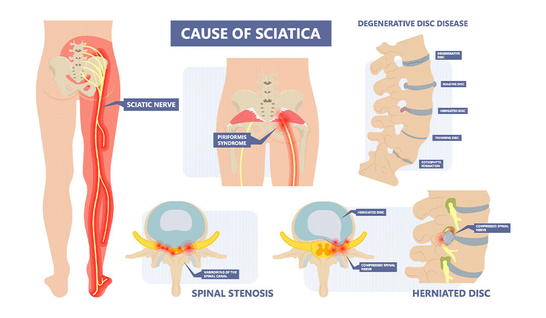
They then moved on to 20 patients with chronic low back pain, many of whom also had pain running down the leg (commonly known as sciatica). The researchers implanted electrodes in all 20; two patients dropped out due to infection at the surgical site.

Among the 18 patients who finished the two-week study, pain ratings improved by an average of 90%. Nearly all of the patients had improvements of at least 80%.

When the electrodes were removed, patients’ back pain came roaring back, according to findings published Aug. 25 in the journal Science Translational Medicine.

“The pain improvement is dramatic — that’s one of the features of this treatment that we find so impressive,” said senior researcher Stephen McMahon, who directs the London Pain Consortium at King’s College London in the United Kingdom.

“Other successful pain therapies more typically find 30% to 50% clinical improvement,” he added.

That said, McMahon cautioned that the study was small and short-term. Further clinical studies will be needed to define the therapy’s effectiveness and how long it lasts, he said.

One of the strengths of this early study is that it “shows directly a powerful inhibition of pain-related signals,” McMahon noted.

Having identified “such a robust mechanism,” he added, it may be possible to use the technique for a range of conditions other than back pain.

The study was funded by Presidio Medical, Inc., of South San Francisco, which is developing the technology.

Danesh said, “I think this is continuing a trend of a technological jump in the use of spinal cord stimulation.”

However, he stressed, no matter what treatments people use for low back pain, some low-tech fundamentals remain key — namely, addressing bad posture habits and muscle strength imbalances.

Sitting all day, and the resulting weakening of the gluteal muscles (in the buttocks), is a big culprit, Danesh noted.

So strengthening those muscles, along with being generally active, is a must.

“You have to be mobile, when you’re in pain and when you’re not,” Danesh said. “Movement is medicine.”

More information

Johns Hopkins University has more on non-surgical treatments for low back pain.

SOURCES: Stephen McMahon, PhD, FMedSci, professor, physiology, and director, London Pain Consortium, King’s College London, U.K.; Houman Danesh, MD, associate professor, anesthesiology, perioperative and pain medicine, Icahn School of Medicine at Mount Sinai, and director, integrative pain management, Mount Sinai Hospital, New York City; Science Translational Medicine, Aug. 25, 2021, online

What to Do If You Wake Up in Pain

What to Do If You Wake Up in Pain

Feeling achy and stiff in the morning? Try these seven techniques to ease into the day.

Nothing is more restorative than a good night’s sleep. You wake up refreshed and ready to take on a new day. Yet, for some people, the early morning hours bring unwelcome neck and back pain.

Movement during the day promotes fluid secretion from your tissues, which in turn keeps your joints mobile. When you’re asleep, however, you’re not moving for an extended period of time, which can cause your joints and muscles to stiffen up.

For some people, morning pain and stiffness go away as the day goes on. For others, the pain lasts all day. If you find yourself waking up constantly in pain, try these seven tips for a pain-free morning:

1. Stretch

When you wake up, do some simple stretches to unlock your joints and muscles. Here are two to try:

  • Knee to Chest Stretch: Remain on your back in your bed, and pull both knees toward you until your feet are flat on the bed. Grab your right knee with both hands and pull toward your chest. Do the same with the left knee. Hold for about 30 seconds. Repeat two to three times.
  • Shoulder/Upper Back Stretch: Stand up and hold your right elbow with your left hand. Stretch your right arm across the front of your body, keeping your arm straight. Do the same with your opposite arm. Hold for 30 seconds, and do two to three repetitions.

2. Change Your Mattress and Pillow

Your morning pain and stiffness could be caused by your bed or pillow. Be sure to choose a pillow that aligns your neck parallel to the mattress so it isn’t bent up or down. As for your mattress, it should be firm, but not too firm.

3. Change Your Sleep Position

Sleeping on your stomach may be the root of your pain. If so, switch to your back and prop up your knees with a pillow to keep your spine in a neutral position. Side-sleepers can try inserting a pillow between their knees.

4. Exercise Regularly

An intense workout just before you slip under the covers is not advisable. However, a brisk walk or relaxing yoga during the day can loosen up your muscles and joints, setting you up for a pain-free morning. Typically, thirty minutes of exercise a day will tire you out so you can fall asleep faster.

5. Stay Hydrated

During the night, you lose water when you sweat, which can lead to dehydration. Dehydration stiffens your muscles and joints as your tissues are deprived of fluids. So instead of going for that cup of morning coffee, drink two glasses of water first to rehydrate your body.

6. Take Vitamin D

Most people don’t get enough vitamin D in their diet. But the vitamin is essential for bone and muscle health. Have your doctor test your vitamin D levels, and if you are deficient, take a vitamin D supplement regularly.

7. Visit a Physical Therapist

Lingering morning pain should be checked out by a physical therapist. Keep a log of where the pain is strongest and which actions seem to aggravate it. Your physical therapist can show you how to adjust your sleeping position to prevent pain and improve your posture. He or she can also recommend the right pillow and mattress for a restful night’s slumber.

Don’t Live With Pain

If you’re experiencing pain and stiffness in the morning, the orthopedic specialists at Comprehensive Orthopaedics can review your symptoms to determine the source of your discomfort.

Tennis Elbow (Lateral Epicondylitis)

Tennis Elbow (Lateral Epicondylitis)

Tennis elbow, or lateral epicondylitis, is a painful condition of the elbow caused by overuse. Not surprisingly, playing tennis or other racquet sports can cause this condition. However, several other sports and activities besides sports can also put you at risk.  Tennis elbow is inflammation or, in some cases, microtearing of the tendons that join the forearm muscles on the outside of the elbow. The forearm muscles and tendons become damaged from overuse — repeating the same motions again and again. This leads to pain and tenderness on the outside of the elbow.There are many treatment options for tennis elbow. In most cases, treatment involves a team approach. Primary doctors, physical therapists and, in some cases, surgeons work together to provide the most effective care.

Anatomy

Your elbow joint is a joint made up of three bones: your upper arm bone (humerus) and the two bones in your forearm (radius and ulna). There are bony bumps at the bottom of the humerus called epicondyles, where several muscles of the forearm begin their course. The bony bump on the outside (lateral side) of the elbow is called the lateral epicondyle.

ECRB muscle and tendon

The ECRB muscle and tendon is usually involved in tennis elbow.
Reproduced and modified from The Body Almanac. © American Academy of Orthopaedic Surgeons, 2003.

Muscles, ligaments, and tendons hold the elbow joint together.

Lateral epicondylitis, or tennis elbow, involves the muscles and tendons of your forearm that are responsible for the extension of your wrist and fingers. Your forearm muscles extend your wrist and fingers. Your forearm tendons — often called extensors — attach the muscles to bone.  The tendon usually involved in tennis elbow is called the Extensor Carpi Radialis Brevis (ECRB).

Cause

Overuse

Recent studies show that tennis elbow is often due to damage to a specific forearm muscle. The extensor carpi radialis brevis (ECRB) muscle helps stabilize the wrist when the elbow is straight. This occurs during a tennis groundstroke, for example. When the ECRB is weakened from overuse, microscopic tears form in the tendon where it attaches to the lateral epicondyle. This leads to inflammation and pain.

The ECRB may also be at increased risk for damage because of its position. As the elbow bends and straightens, the muscle rubs against bony bumps. This can cause gradual wear and tear of the muscle over time.

Activities

Athletes are not the only people who get tennis elbow. Many people with tennis elbow participate in work or recreational activities that require repetitive and vigorous use of the forearm muscle or repetitive extension of the wrist and hand.

Painters, plumbers, and carpenters are particularly prone to developing tennis elbow. Studies have shown that auto workers, cooks, and even butchers get tennis elbow more often than the rest of the population. It is thought that the repetition and weight lifting required in these occupations leads to injury.

Tennis player

Playing tennis is a possible cause of tennis elbow, but other activities can also put you at risk.

Thinkstock © 2015.

Age

Most people who get tennis elbow are between the ages of 30 and 50, although anyone can get tennis elbow if they have the risk factors. In racquet sports like tennis, improper stroke technique and improper equipment may be risk factors.

Unknown

Lateral epicondylitis can occur without any recognized repetitive injury. This occurrence is called “idiopathic” or of an unknown cause.

Symptoms

The symptoms of tennis elbow develop gradually. In most cases, the pain begins as mild and slowly worsens over weeks and months. There is usually no specific injury associated with the start of symptoms.

Common signs and symptoms of tennis elbow include:

  • Pain or burning on the outer part of your elbow
  • Weak grip strength
  • Sometimes, pain at night

The symptoms are often worsened with forearm activity, such as holding a racquet, turning a wrench, or shaking hands. Your dominant arm is most often affected; however, both arms can be affected.

Location of pain in lateral epicondylitis.

Location of pain in lateral epicondylitis.

Doctor Examination

Your doctor will consider many factors in making a diagnosis. These include how your symptoms developed, any occupational risk factors, and recreational sports participation.

Your doctor will talk to you about what activities cause symptoms and where on your arm the symptoms occur. Be sure to tell your doctor if you have ever injured your elbow. If you have a history of rheumatoid arthritis or nerve disease, tell your doctor.

During the examination, your doctor will use a variety of tests to pinpoint the diagnosis. For example, your doctor may ask you to try to straighten your wrist and fingers against resistance with your arm fully straight to see if this causes pain. If the tests are positive, it tells your doctor that those muscles may not be healthy.

doctor pressing lateral epicondyle

During the exam, your doctor will apply gentle pressure to the lateral epicondyle, checking for pain and tenderness.

Tests

Your doctor may recommend additional tests to rule out other causes of your problem.

  • X-rays. These tests provide clear images of dense structures, such as bone. They may be taken to rule out arthritis of the elbow.
  • Magnetic resonance imaging (MRI) scan.  MRI provides images of the body’s soft tissues, including muscles and tendons. An MRI scan may be ordered to determine the extent of damage in the tendon or to rule out other injuries. If your doctor thinks your symptoms might be related to a neck problem, he or she may order an MRI scan of the neck to see if you have a herniated disk or arthritic changes in your neck. Both of these conditions can produce arm pain.
  • Electromyography (EMG). Your doctor may order an EMG to rule out nerve compression. Many nerves travel around the elbow, and the symptoms of nerve compression are similar to those of tennis elbow.

Treatment

Nonsurgical Treatment

Approximately 80% to 95% of patients have success with nonsurgical treatment.

Rest. The first step toward recovery is to give your arm proper rest. This means that you will have to stop or decrease participation in sports, heavy work activities, and other activities that cause painful symptoms for several weeks.

Medications. Acetaminophen or anti-inflammatory medications (such as ibuprofen) may be taken to help reduce pain and swelling

Physical therapy. Specific exercises are helpful for strengthening the muscles of the forearm. Your therapist may also perform ultrasound, ice massage, or muscle-stimulation techniques to improve muscle healing.

Wrist stretching exercise

Wrist stretching exercise with elbow extended.

Brace. Using a brace centered over the back of your forearm may also help relieve symptoms of tennis elbow. This can reduce symptoms by resting the muscles and tendons.

Counterforce brace

Counterforce brace.

Steroid injections. Steroids, such as cortisone, are very effective anti-inflammatory medicines. Your doctor may decide to inject the painful area around your lateral epicondyle with a steroid to relieve your symptoms.

Platelet-rich plasma. Platelet-rich plasma (PRP) is a biological treatment designed to improve the biologic environment of the tissue. This involves obtaining a small sample of blood from the arm and centrifuging it (spinning it) to obtain platelets from the solution.  Platelets are known for their high concentration of growth factors, which can be injected into the affected area. While some studies about the effectiveness of PRP have been inconclusive, others have shown promising results.

PRP injection

An injection of PRP is used to treat tennis elbow.
Courtesy of Allan K. Mishra, MD, Menlo Park, CA.

Extracorporeal shock wave therapy. Shock wave therapy sends sound waves to the elbow. These sound waves create “microtrauma” that promotes the body’s natural healing processes. Shock wave therapy is considered experimental by many doctors, but some sources show it can be effective.

Equipment check. If you participate in a racquet sport, your doctor may encourage you to have your equipment checked for proper fit. Stiffer racquets and looser-strung racquets often can reduce the stress on the forearm, which means that the forearm muscles do not have to work as hard. If you use an oversized racquet, changing to a smaller head may help prevent symptoms from recurring.

Surgical Treatment

If your symptoms do not respond after 6 to 12 months of nonsurgical treatments, your doctor may recommend surgery.

Most surgical procedures for tennis elbow involve removing diseased muscle and reattaching healthy muscle back to bone.

The right surgical approach for you will depend on a range of factors. These include the scope of your injury, your general health, and your personal needs. Talk with your doctor about the options. Discuss the results your doctor has had, and any risks associated with each procedure.

Open surgery. The most common approach to tennis elbow repair is open surgery. This involves making an incision over the elbow.

Open surgery is usually performed as an outpatient surgery. It rarely requires an overnight stay at the hospital.

Arthroscopic surgery. Tennis elbow can also be repaired using miniature instruments and small incisions. Like open surgery, this is a same-day or outpatient procedure.

Surgical risks. As with any surgery, there are risks with tennis elbow surgery. The most common things to consider include:

  • Infection
  • Nerve and blood vessel damage
  • Possible prolonged rehabilitation
  • Loss of strength
  • Loss of flexibility
  • The need for further surgery

Rehabilitation. Following surgery, your arm may be immobilized temporarily with a splint. About 1 week later, the sutures and splint are removed.

After the splint is removed, exercises are started to stretch the elbow and restore flexibility. Light, gradual strengthening exercises are started about 2 months after surgery.

Your doctor will tell you when you can return to athletic activity. This is usually 4 to 6 months after surgery. Tennis elbow surgery is considered successful in 80% to 90% of patients. However, it is not uncommon to see a loss of strength.

Get Relief for Hip Pain at Any Age

Get Relief for Hip Pain at Any Age

Arthritis isn’t the only cause of hip pain. Find out which condition may be causing your discomfort — and how to treat it.

Hip pain doesn’t only affect older adults — stiffness and pain in this joint can strike younger people, too. Although osteoarthritis ranks high on the list of causes of hip pain, the discomfort can result from overuse or injuries at any age.

In most cases, the pain radiates from the side of the hip or groin. It can also be felt in the buttocks. The pain may worsen when you stand, walk, sit for long periods, or twist the hips. If your pain persists, see an orthopedist for a correct diagnosis and treatment.

What Causes Hip Pain?

To determine the exact cause of your hip pain, your doctor will analyze your movements, review your symptoms, and order imaging tests, such as an X-ray or MRI. Those details should provide a picture of what’s happening in your hip.

Common causes of hip pain include:

OsteoarthritisWhen the cartilage cushioning the hip joint wears down, it can lead to pain and stiffness. Mostly the result of advanced age, osteoarthritis may also be due to a fracture or infection of the hip joint.

Bursitis. The bursae, or the fluid-sacs between the bones and soft tissue of the hip joint, sometimes become inflamed when the hip is stressed from overuse. That’s why bursitis is especially common among runners.

Labral Hip Tear. The hip joint is formed by the femur (the ball) and the pelvic acetabulum (the socket). A ring of cartilage — the acetabular labrum — surrounds the joint. An injury or a malformed hip (known as hip dysplasia) may cause the femoral head to grind against the acetabulum, eventually rupturing the labrum.

Hip Impingement. A hip impingement develops when the bones of the hip joint rub against each other, causing significant pain. The most likely cause is an ill-fit between the ball and socket of the hip joint, which can also lead to cartilage damage.

Fracture. As we age, our bones weaken and become more susceptible to fracture. If you fall and feel sharp, sudden pain, seek immediate medical attention. A blood clot in the leg can form following a hip fracture, which makes it especially important to act fast.

Osteonecrosis. Osteonecrosis, or avascular necrosis, is a breakdown of the hip bone because blood fails to penetrate the bones. Over time, the cartilage and bones wear away, leading to severe bone loss. In most cases, a definitive cause is not determined, although joint trauma, excessive steroid use, and certain cancer treatments may put a person at greater risk.

Snapping Hip Syndrome. Another rare condition, snapping hip syndrome, is characterized by a snapping sound or feeling in the hip, particularly when you walk or rise from a chair. Dancers and athletes are prone to this condition, which is usually painless.

Treatment for Hip Pain

Once you’ve been diagnosed, your doctor will recommend a treatment plan, which can range from conservative therapy to surgery. If the pain is due to overuse, a few days of rest can heal the strained joint or tendon. For arthritis, pain medication and physical therapy can help heal the pain and encourage freedom of movement.

Some conditions, however, may require surgery. If a labrum tear or impingement doesn’t respond to conservative treatments like physical therapy, arthroscopic surgery can repair the damaged cartilage. Therapies for bursitis typically involve physical therapy and anti-inflammatory medications. In severe cases of bursitis, surgery may be an option to drain the bursa or remove it altogether.

If the damage to the hip is substantial, either due to advanced arthritis or a fracture, hip replacement surgery is available. After surgery, intensive physical therapy will be needed to get you acclimated to your new joint.

If your hip pain doesn’t fade in a couple of days, it’s time to see a specialist at CompOrtho. Using the latest technology, we’ll diagnose your condition and recommend a customized treatment plan. Contact us today for a consultation.

More Than Half of Americans Plagued by Back, Leg Pain

More Than Half of Americans Plagued by Back, Leg Pain

There’s much Americans may disagree on, but many share one thing in common: chronic pain.

More than half of U.S. adults suffer from pain, with backs and legs the most common sources, according to researchers from the U.S. Centers for Disease Control and Prevention’s National Center for Health Statistics (NCHS).

Overall, the investigators found that nearly 59% of American men and women were saddled with pain.

“Pain is one of the most common presenting complaints to a doctor’s office,” said Dr. Yili Huang, director of the Pain Management Center at Northwell Health’s Phelps Hospital, in Sleepy Hollow, N.Y.

“It is often the body’s warning sign that something may be wrong. Pain is a symptom and not a disease, so when experiencing new pain, it is important to seek medical advice to help diagnose the cause and to ensure that it is not an emergent or urgent medical condition,” said Huang, who was not involved in the research.

Using 2019 data from the National Health Interview Survey, the NCHS researchers found that 39% of adults had back pain, 37% had hip, knee or foot pain, and nearly one-third had hand, arm or shoulder pain in the past three months. About one in 10 suffered from toothaches.

The goal of the survey was not to draw conclusions from the data, but to provide the groundwork for further analyses, the researchers said.

“Given what we know about the short- and long-term effects of pain, timely, up-to-date national estimates of location-specific pain are an important step in understanding the burden of pain on U.S. adults,” said lead author Jacqueline Lucas, a health statistician/epidemiologist at the NCHS in Hyattsville, Md.

The odds of experiencing pain were related to economics and age, according to the report published July 29 in an NCHS Data Brief.

Those 65 and older, women, white adults and those with incomes below the federal poverty level ($25,750 for family of four in 2019) were most likely to have back pain and lower limb pain, as well as hand, arm or shoulder pain, the report noted.

Among those least likely to experience pain were those aged 18 to 29, men, Asian adults and those with an income 200% of the federal poverty level or above.

Huang said chronic pain is often musculoskeletal and associated with degenerative wear-and-tear, often due to a physically demanding job or aging.

“Women, especially those who are postmenopausal, have a higher incidence of musculoskeletal pain, possibly because of hormonal differences, although the association between hormones and lower back pain is not well understood,” Huang said.

Socioeconomic factors and health are inherently linked, he added.

“It is well-established that pain is associated with social-economic status. Part of that is likely because those who have chronic lower back pain often have physically demanding work. Also, studies have shown a strong association with chronic back, leg and arm pain, and patients with the lowest educational levels and blue-collar workers, likely because of the nature of the work,” Huang said.

Most chronic musculoskeletal pain is probably caused by degeneration and age. Also, “being overweight can lead to added stress to joints of the back and extremities as well, which can lead to increased degeneration,” he noted.

Most people will develop chronic degenerative pain sometime in their lives. “The key to treatment is to focus on quality of life and function, not just the pain,” Huang said.

That doesn’t mean just living with it, however.

“Activities that help us adapt to whatever is causing the pain can be helpful. That can be physical therapy, like exercises, or psychological therapy, like biofeedback or meditation, or medications,” Huang said. “It often makes sense to find a medical professional that is able to help diagnose the source of pain and work with you to find safe options to treat it.”

More information

For more on pain, head to the U.S. National Library of Medicine.

SOURCES: Jacqueline Lucas, MPH, health statistician/epidemiologist, U.S. Centers for Disease Control and Prevention, National Center for Health Statistics, Hyattsville, Md.; Yili Huang, DO, director, Pain Management Center, Northwell Health’s Phelps Hospital, Sleepy Hollow, N.Y.; NCHS Data Brief, July 29, 2021

Copyright ©2021 HealthDay. All rights reserved.
Call Now Button