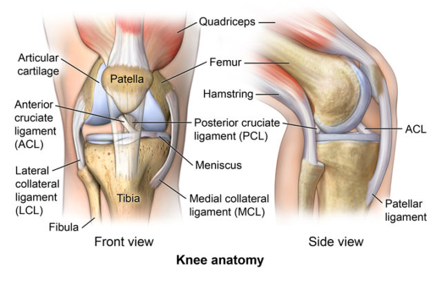
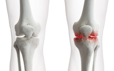
The knee is a vulnerable joint that bears a great deal of stress from everyday activities such as lifting and kneeling, and from high-impact activities such as jogging and aerobics.
The knee is formed by the following parts:
- Tibia. Shin bone or larger bone of the lower leg
- Fibula. Smaller of the 2 lower leg bones
- Femur. Thighbone or upper leg bone
- Patella. Kneecap
Each bone end is covered with a layer of cartilage that absorbs shock and protects the knee. Basically, the knee is 2 long leg bones held together by muscles, ligaments, and tendons.
There are 2 groups of muscles involved in the knee, including the quadriceps muscles (located on the front of the thighs), which straighten the legs, and the hamstring muscles (located on the back of the thighs), which bend the leg at the knee.
Tendons are tough cords of tissue that connect muscles to bones. Ligaments are elastic bands of tissue that connect bone to bone. Some ligaments on the knee provide stability and protection of the joints, while other ligaments limit forward and backward movement of the tibia (shin bone).


